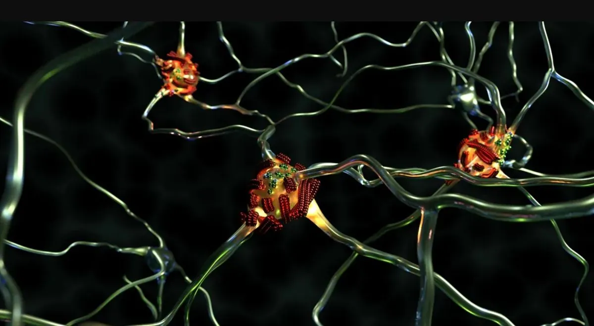
For the first time, researchers have traced the formation of amyloids associated with Alzheimer's disease with unprecedented accuracy
Kyiv • UNN
Empa researchers visualized the process of amyloid β protein accumulation in the brain associated with Alzheimer's disease. The observation lasted 250 hours and showed how second-generation fibrils are formed.

Empa researchers have recently been able to visualize the “abnormal” accumulation of amyloid β proteins in the brain, a process associated with Alzheimer's disease. Scientists hope that the result will open up new methods of diagnosing the disease.
Writes UNN with reference to Science Daily.
Alzheimer's disease affects more than 55 million people worldwide. Treatment of such disorders is still one of the biggest challenges facing modern medicine. The accumulation of amyloid plaques in the brain plays a significant role in the development of the disease.
Misfolded proteins stick together to form fiber-like structures. However, it was not yet entirely clear how these fibrils are formed.
Recently, a team led by Empa researcher Peter Nirmalraj from the Empa Transport at Nanoscale Interfaces lab and scientists from the University of Limerick in Ireland were able to show how this process works using a particularly powerful visualization technique.
ExplanationExplanation
The focus is on the processes observed on the surface of amyloid fibrils, i.e. structures in the brain formed by oligomers - soluble proteins that are highly toxic to neurons and assemble into dense fibers. Scientists have identified a special subtype of superproliferator proteins among them.
For reference
Aβ42 is considered to be one of the main factors that trigger Alzheimer's disease.
Incorrectly folded proteins lead to the formation of new oligomers on the surface of existing fibrils during secondary nucleation (when mature fibrils catalyze the formation of new oligomers).
Aβ is formed from a larger transmembrane precursor protein (beta-amyloid precursor, PBA) with a molecular weight of 100-140 kDa, which performs trophic and protective functions.
According to the “amyloid hypothesis,” beta-amyloid forms neurotoxic soluble oligomers for reasons that are not fully understood. According to numerous studies, beta-amyloid aggregates, including oligomers, have a detrimental effect on nerve cells, disrupting their normal functioning. This creates conditions for the development of Alzheimer's disease.
What the new observation achieves
The mechanism of how the protein building blocks come together to form second-generation fibrils, as well as their shape and structure, were previously unclear.

Recent imaging brings us one step closer to a better understanding of how these proteins are distributed in Alzheimer's brain tissue.
Scientists develop nanoparticles to fight obesity14.10.24, 12:45 • 11771 view
The researchers were able to observe the process of fibril formation in real time, from the first moments to the next 250 hours. This was made possible by a new technique: proteins are analyzed unchanged in saline, which is much closer to the natural conditions of the human body.
“This work brings us one step closer to better understanding how these proteins are distributed in Alzheimer's brain tissue,” says Nirmalraj, an Empa researcher. He hopes this will eventually lead to new ways of diagnosing the disease.
Scientists discover new sign of early stage of Alzheimer's disease28.10.24, 17:27 • 16022 views